


Foreign Material Inspection and Detection Methods
X-ray non-destructive testing (NDT) is a versatile tool used across various industries, primarily for detecting foreign contaminants. This section outlines the procedures and key features of foreign material inspection, using the food manufacturing process as a primary example.
In the food industry, consumer safety is the highest priority, often requiring 100% inspection of products. The HACCP (Hazard Analysis and Critical Control Point) food safety standard categorizes hazards into three types:
- B (Biological): Contamination by microorganisms
- C (Chemical): Chemical substances
- P (Physical): Physical foreign materials/li>
This principle extends beyond food production. Products packaged in metal cans or foil pouches cannot be inspected with metal detectors. Furthermore, contaminants like stones do not trigger metal detectors. In these cases, X-ray inspection is the only viable solution for quality assurance.
Of course, non-destructive inspection by X-ray is also used in manufacturing processes other than food. Items in metal cases and cans can only be inspected by X-rays, and stones and the like do not react to metal detectors, so X-ray inspection is required to find them.
Alternative inspection methods, such as Fourier Transform Infrared Spectrophotometry (FT-IR), are also used to identify foreign materials. FT-IR analyzes the material by irradiating the sample with infrared rays and identifying the absorbed wavelengths. Unlike X-ray inspection, which produces an image of the internal structure, FT-IR identifies the material composition based on its infrared absorption spectrum. This is particularly useful for analyzing liquids in bottles or screening baggage at airports to distinguish between water and hazardous chemicals.
However, when identifying contaminants based on shape or density inside a product, X-ray inspection is superior. Energy-dispersive X-ray spectroscopy or Microfocus X-ray CT systems are employed to visualize and identify foreign materials. By comparing the captured image against a database of known contaminants (such as metal fragments, bone, or dense fibers), automated systems can detect and classify contamination.
For detailed internal analysis, Microfocus X-ray CT allows for non-destructive cross-sectional imaging. While neutron imaging is an option for materials that are transparent to X-rays, the equipment is typically large and complex. Therefore, X-ray CT remains the most practical choice for industrial defect and contaminant detection.
Basics of Image Processing
Raw X-ray images often require processing to extract meaningful data for automated inspection.
Binarization
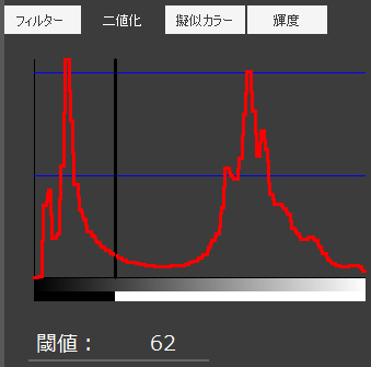
Binarization converts a grayscale image into black and white based on a specific brightness threshold. Areas above the threshold become white (value 255 in 8-bit scale), and areas below become black (value 0). This process separates the target object from the background, allowing for area calculation and shape extraction.
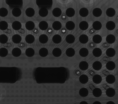

Example of Binarization (left: original, right: binarized)
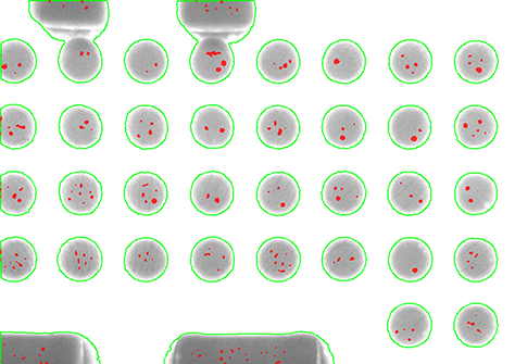
Contour Extraction
Contour extraction is often applied after binarization. By tracing the boundary pixels between the black and white regions, the system can define the outline of the object. This allows the software to measure the sample's size, shape, and count.
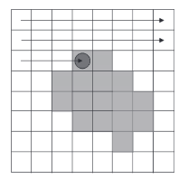
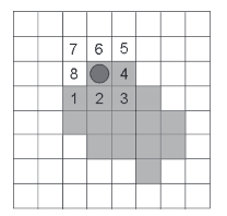
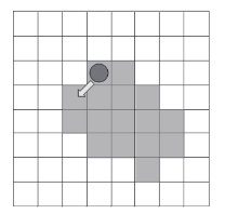
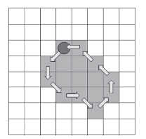
Contour Detection Method
Edge Detection
For grayscale images that are not binarized, edge detection is used. Unlike contour extraction which looks for a simple black/white boundary, edge detection identifies areas where brightness changes rapidly (a high brightness gradient). This is essential for detecting features within an object that has varying densities.
Gradation Correction
To improve detection accuracy, gradation correction techniques such as Histogram Equalization (to increase contrast) or Gamma Correction (to adjust brightness non-linearly) are frequently applied.

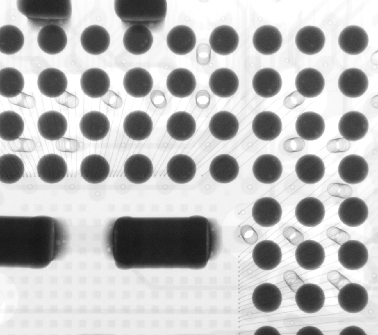
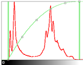
Image-to-Image Operations
Processing techniques can also involve combining multiple images, known as image-to-image operations.
- Addition/Superposition: Combining images to reduce noise or enhance faint details.
- Subtraction: Comparing a reference image with a current image to highlight differences. In subtraction, unchanged areas result in a brightness value near zero (black), while changes (such as foreign materials or defects) appear clearly.

Example of Subtraction (Left: 1st image, Center: 2nd image, Right: subtracted)
Pseudo-Color Imaging
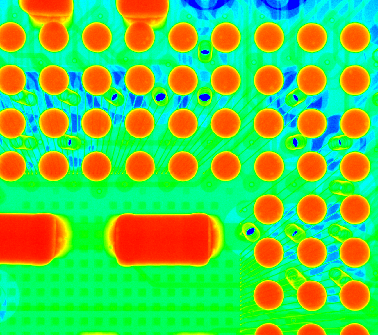
Human eyes typically struggle to distinguish subtle differences in grayscale levels. Pseudo-color processing maps brightness values to specific colors to make these differences visible.
For example, in a thermal image (as shown below), hotter areas might be mapped to red or white, while colder areas appear blue. Similarly, in X-ray inspection, variations in density that are difficult to see in grayscale can be highlighted by assigning different colors to specific intensity ranges.
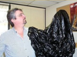
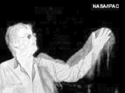
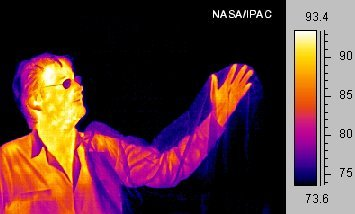
Pseudo-Color Example (Public Domain)
Thus, image processing combines various techniques depending on the information to be extracted and how it needs to be presented.
Related Technical Articles
- Basics and Principles of Computed Tomography (CT)
- How to Acquire High-Quality Computed Tomography (CT) Images - X-ray NDT series (1)
- A Guide to X-ray CT Images: Formats, Viewing, and Applications - X-ray NDT series (2)
- FAQ: What is the tube voltage or acceleration voltage necessary for X-ray inspection systems?
- Non-Destructive Testing: Types and Applications
Recommended products
Matsusada Precision's X-ray non-destructive inspection system can take high-definition and High-resolution images with its unique microfocus X-ray technology.


Reference (Japanese site)
- Japanese source page 「X線非破壊検査シリーズ③ 自動検査と画像処理」
(https://www.matsusada.co.jp/column/x-xct3.html) - 岐阜県保健環境研究所「食品中の異物検査法の構築」
(https://www.health.rd.pref.gifu.lg.jp/kankoubutu/shohou/26/2018_26_foreign%20matter.pdf) - 広島県「非破壊検査・異物分析」
(https://www.pref.hiroshima.lg.jp/uploaded/attachment/119898.pdf, dead link) - 非破壊検査と画像処理
(https://www.jstage.jst.go.jp/article/tetsutohagane1955/70/9/70_9_1000/_pdf/-char/ja)