


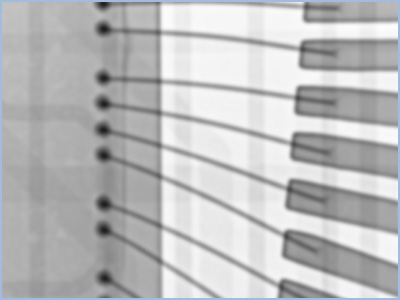
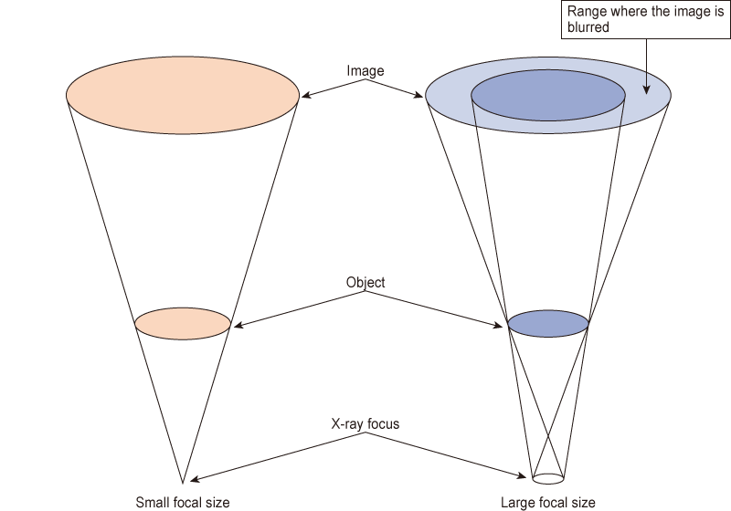
The focal spot size of an X-ray source directly influences image sharpness. When inspecting components such as ICs (as shown in the comparison), a smaller focal spot size significantly reduces geometric blur, resulting in a clearer, higher-resolution image.
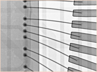
Conventional X-ray sources, such as those often used in general medical radiography, typically have focal spot sizes measured in millimeters. While effective for larger structures, these sources naturally produce geometric unsharpness (blur) when high magnification is required.
In contrast, microfocus X-ray sources feature focal spot sizes measured in micrometers (microns). By minimizing the source size, these systems achieve superior spatial resolution. They are essential for non-destructive testing (NDT) applications where high magnification and detailed imaging are critical.
When relying on visual data for quality assurance, image clarity is paramount. High-resolution microfocus X-ray systems allow operators to clearly identify minute defects and abnormalities in electronics, precision mechanical parts, and advanced materials.
For more information about these products, see X-Ray inspection systems / X-Ray CT.
In addition, our experienced staff will answer any inquiries you may have regarding intended use and desired specifications. Please contact us via the Contact pages. Our staff will be happy to answer all of your questions.
Related Technical Articles
Recommended products
Matsusada Precision's X-ray non-destructive inspection system can take high-definition and High-resolution images with its unique microfocus X-ray technology.


